

Mammogram in Singapore

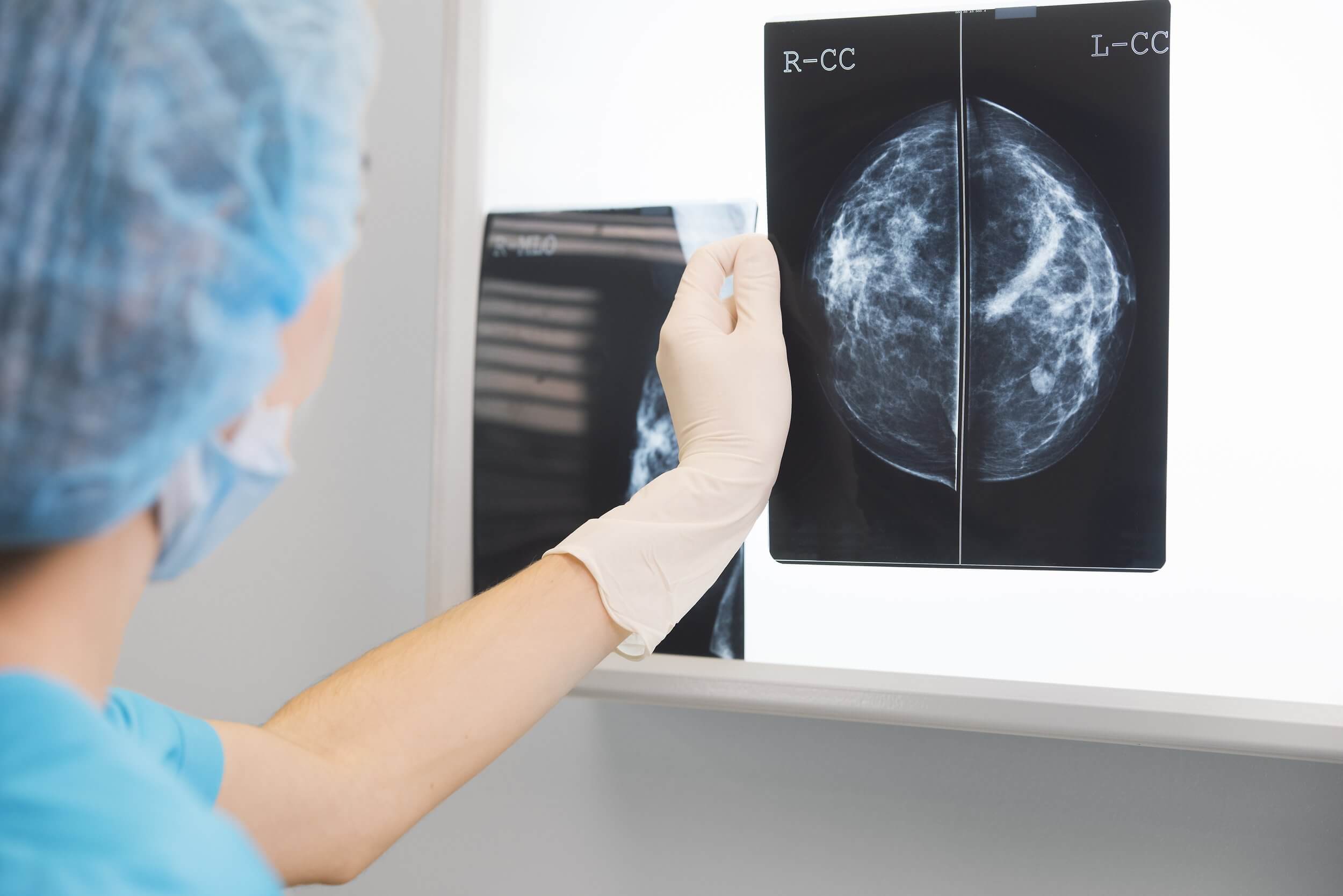
A mammogram uses low-dose X-rays to produce clear, detailed images of the breast, and can detect certain breast changes before they can be felt or cause noticeable symptoms. It is commonly used for breast cancer screening and may also form part of a diagnostic breast assessment when further investigation is needed.
At Solis Breast Care & Surgery Centre, 3D mammography is routinely performed together with breast ultrasound. Your imaging findings can then be considered alongside your clinical history and breast examination by a breast specialist, helping determine whether further assessment is needed and what the next steps should be.

When is a Mammogram Recommended?
Breast cancer may not cause any noticeable symptoms in its early stages. Regular mammogram screening helps identify changes in the breast tissue early, sometimes before a lump can be felt or other symptoms develop. Some breast cancers detected through screening may be diagnosed at an early stage, including Stage 1 breast cancer, allowing timely assessment and appropriate management.
If an abnormality is detected during a mammogram, your breast specialist will guide you through any further assessment and explain the recommended next steps. Where appropriate, they will discuss the most suitable treatment options and develop a personalised care plan based on your individual condition.
For women at average risk, mammography may be considered from age 40, with the frequency depending on age and individual circumstances. Women aged 40 to 49 should discuss the potential benefits and limitations of screening with their doctor. From age 50, mammography every two years is generally recommended. Screening recommendations may differ for women with additional breast cancer risk factors.
Mammograms for Dense Breast Tissue
Dense breast tissue is common, particularly among Asian women. It simply means that the breasts contain more fibrous and glandular tissue than fatty tissue and is a normal variation in breast composition. While dense breast tissue is not a cause for concern on its own, it can make some breast abnormalities more difficult to detect on a mammogram, as both dense tissue and certain abnormalities appear white on the images.
In addition to affecting how breast tissue appears on a mammogram, dense breast tissue is associated with a higher risk of breast cancer. For this reason, your breast specialist will consider your breast density, together with your clinical history, examination findings and other individual risk factors, when recommending the most appropriate assessment or follow-up plan for you.
How 3D Mammography Supports Breast Assessment
Mammography technology has advanced significantly, providing breast specialists with more ways to assess breast tissue. The two main types of mammography are:
2D Mammography
Uses low-dose X-rays to produce two-dimensional images of the breast, typically captured in two standard views of each breast. It is the conventional mammography technique used to detect breast abnormalities.
3D Mammography (Breast Tomosynthesis)
3D Mammography and Breast Ultrasound
Mammogram vs Ultrasound
Mammography and breast ultrasound are different imaging techniques that provide complementary information. They may be used together depending on your individual circumstances.
Mammogram | Breast Ultrasound | |
How it works | Uses low-dose X-rays to create images of the breast | Uses sound waves to produce images of breast tissue |
Common uses | Breast cancer screening and assessment of abnormalities | Assessment of breast abnormalities, including lumps or areas of concern; often used alongside mammography |
What it can detect | Masses, asymmetries, and some microcalcifications | Can help distinguish cysts from solid masses and assess lymph nodes and other areas of concern |
Dense breasts | Some abnormalities may be more difficult to detect in women with dense breast tissue | Provides additional information and complements mammography, including in women with dense breast tissue |
Radiation | Uses a low dose of radiation | Does not use ionising radiation |
What Can You Expect During Your Mammogram?
If it’s your first mammogram, it’s natural to feel a little unsure about what to expect. The procedure is straightforward and usually takes only a short time. Here’s what you can expect during your visit.
Before Your Appointment
On the day of your mammogram, avoid applying deodorant, powder, perfume or lotion to your breasts or underarms, as these products can affect the clarity of the images. Wear a two-piece outfit or clothing that is easy to remove from the waist up. If you are pregnant or think you may be pregnant, let your breast specialist or radiographer know before the scan.
During the Mammogram
You will be asked to undress from the waist up. Each breast will be positioned between two plates and gently compressed for a few seconds while the X-ray images are taken. The compression may feel slightly uncomfortable, but it is brief and helps produce clear images and reduces the chance that breast tissue will be obscured.
How long it takes:
A mammogram appointment usually takes around 15 to 30 minutes.
After Your Appointment
You can return to your usual daily activities straight away. Your mammogram images will be reviewed by a radiologist, and your results will be communicated according to the arrangements made with your healthcare provider.
If you require further assessment following a mammogram or have concerns about your breast health, our breast specialists can provide personalised advice and guide you through the next steps. Book a consultation with Solis Breast Care & Surgery Centre.
Understanding Your Mammogram Results
Waiting for your mammogram results can feel worrying, especially if it’s your first imaging test. It is important to remember that being called back for further assessment does not necessarily mean breast cancer has been found. In many cases, additional tests are simply recommended to gather more information or to take a closer look at an area seen on the mammogram.
There are many possible reasons for an abnormal or inconclusive result, including common benign breast conditions, overlapping breast tissue or cysts. Your breast specialist will explain your results clearly, answer any questions you may have, and discuss what they mean for your individual situation.
If further assessment is recommended, it may involve additional mammogram views, a breast ultrasound, an MRI or a biopsy. These investigations provide more detailed information and help your specialist determine whether any further treatment or follow-up is needed. Throughout the process, the care team will guide you through each step and ensure you understand the recommendations and options available to you.
Important Considerations Before A Mammogram
Mammography is a safe, well-established and widely used breast imaging test. While it is suitable for many women, there are a few important considerations to keep in mind before your appointment:
- Mammograms use a very small amount of radiation, and the benefits of screening generally outweigh the risks for women who are recommended to undergo screening.
- Breast compression is necessary to obtain clear images. It may feel uncomfortable for a few seconds, but the discomfort is usually brief and temporary.
- Like any screening test, mammograms are not perfect. Occasionally, they may identify an area that requires further assessment even though it is not cancer (a false positive), or they may not detect every breast abnormality (a false negative).
- Women under 40, women who are pregnant or breastfeeding and women with breast implants may require an individualised imaging approach depending on their circumstances. Your breast specialist will advise you on the most appropriate investigation.
- If you have symptoms such as a new breast lump, nipple discharge, persistent breast pain, or new and persistent changes in the appearance of your breast, you may require a diagnostic breast assessment rather than a routine screening mammogram.
How Much Does A Mammogram Cost, and Is It Covered By Insurance?
The cost of a mammogram varies between providers and depends on whether you need a routine screening or additional imaging, such as extra views or an ultrasound.
MediSave may be used for eligible mammogram screening and certain outpatient diagnostic scans, subject to prevailing withdrawal limits and eligibility requirements. The current outpatient scan limit is up to $600 per patient per year. Insurance coverage also varies by policy, so if you’re considering a mammogram, it’s worth checking with your insurer beforehand to understand whether your plan covers screening, diagnostic imaging, or both.1
Supporting You Beyond Your Mammogram
A mammogram is often the first step in assessing your breast health. If further assessment is needed, Solis Breast Care & Surgery Centre works closely with Luma Women’s Imaging Centre to ensure your diagnostic imaging, specialist consultation and follow-up care are coordinated seamlessly. From diagnosis through to surgical treatment, our integrated approach provides continuity, clarity and reassurance at every stage of your care.
Dedicated diagnostic imaging
Access to breast imaging, including mammography, ultrasound and MRI where appropriate.
Specialist assessment
Mammogram findings are reviewed alongside your clinical history and breast examination to build a complete understanding of your breast health.
Coordinated follow-up
If additional investigations are recommended, such as further imaging or a biopsy, your specialist will explain the findings and guide you through the next steps.
Continuity of care
Where treatment is required, our multidisciplinary team can continue to support you through surgical treatment and follow-up care.
Frequently Asked Questions
At what age should women in Singapore start regular mammogram screening, and how do I choose a clinic?
For women at average risk, mammography may be considered from age 40, with the frequency depending on age and individual circumstances. Women aged 40 to 49 should discuss the potential benefits and limitations of screening with their doctor. From age 50, mammography every two years is generally recommended. Screening recommendations may differ for women with additional breast cancer risk factors.
When choosing where to have your mammogram, you may wish to consider a centre that provides comprehensive breast assessment, including appropriate imaging, specialist review, and access to further investigations, such as a biopsy, as well as surgical treatment if needed.
Can I have a mammogram if I don’t have any breast symptoms?
Yes. Mammograms may be used for routine breast cancer screening in women who do not have breast symptoms, depending on their age and individual risk. If you have a new breast symptom or breast concern, however, you may require a diagnostic breast assessment rather than routine screening.
What should I consider when choosing a mammogram screening clinic in Singapore?
Beyond the mammogram itself, consider whether the clinic provides access to specialist review and a clear pathway for further assessment if an abnormality is identified. Knowing who will explain your results and coordinate any additional imaging, biopsy, or treatment can help ensure a more seamless care experience.
3D mammogram versus 2D mammogram: Which is more accurate for breast cancer detection in Singapore?
Both 2D mammography and 3D mammography (breast tomosynthesis) are effective breast imaging techniques. Compared with 2D mammography, 3D mammography provides more detailed images of the breast and can improve the detection and assessment of certain breast abnormalities, particularly in women with dense breast tissue. However, the most appropriate imaging approach depends on your age, breast density, clinical history and the reason for the examination. Your breast specialist or radiologist will recommend the most suitable option based on your individual needs.
Can a mammogram reliably detect stage 1 breast cancer?
A mammogram can detect some small breast abnormalities before they can be felt or seen, including cancers that may later be confirmed to be at an early stage. However, a mammogram alone cannot determine the stage of a cancer. If an abnormality is detected, further assessment, such as additional imaging and/or a biopsy, may be recommended to confirm whether cancer is present and, if so, determine its stage.
Key Takeaway
A mammogram is an important part of breast imaging and may be used for both screening and diagnostic assessment.
The most appropriate approach depends on your age, breast density, personal and family history, symptoms and other individual risk factors. Your breast specialist will recommend the most suitable assessment based on your needs.
If your mammogram identifies an area that requires further assessment, our breast specialists can explain the findings and guide you through the next steps.
Book a Consultation
At Solis Breast Care & Surgery Centre, we believe breast care begins with a thorough diagnosis. By combining evidence-based medicine with compassionate, patient-centred care, we provide the expertise, support, and guidance you need at every stage of your journey.
Every patient is treated with empathy, respect, and personalised attention, because we know that every breast concern and every patient is unique.
Solis Breast Care & Surgery Centre @ The Paragon (Orchard Road)
290 Orchard Road, Lobby C, #09-30/31, Singapore 238859
Nearest MRT: Orchard Station (North-South Line / TEL Line)
Tel: +65 6994 5499
Solis Breast Care & Surgery Centre @ Mt Elizabeth Hospital (Orchard)
3 Mount Elizabeth, #13-01 Medical Centre, Singapore 228510
Nearest MRT: Orchard Station (North-South Line / TEL Line)
Tel: +65 6994 5477
Solis Breast Care & Surgery Centre @ Royal Square Medical Centre (Novena)
101 Irrawaddy Road, #12-01/02, Singapore 329565
Nearest MRT: Novena Station (North-South Line)
Tel: +65 6262 3202