Why Early Detection for Breast Cancer is Important & Ways to Prevent It

Breast cancer is the most common cancer among women in Singapore with over 2,000 women diagnosed each year. According to the Singapore Cancer Registry Annual Report 2018, the incidences of breast cancer are highest in women aged between 40 and 69 years old. What are some of the symptoms and risk factors, and how can we prevent breast cancer? The most common type of breast cancer originates from the cells lining the milk ducts and glands[1]. When abnormal cells are detected in the milk ducts and have not spread to other parts of the breast or the rest of the body, this is termed as Ductal Carcinoma In-Situ (DCIS). Patients with DCIS have a greater chance of recovering. Breast cancer may also begin in the glandular tissue called lobules or in other cells or tissue within the breast. The importance of early detection and regular breast cancer screening Going for regular breast cancer screening is important because it catches breast changes early before symptoms (such as a lump that can be felt) develop. Breast cancer is most treatable when it is detected and diagnosed at an early stage. Identifying the disease during its initial growth can mean that the required treatments are simpler and more effective. The earlier the breast cancer is detected, the smaller the tumour may be and the less likely it would have spread to other parts of your body or the lymph nodes. Breast cancer symptoms Symptoms may vary for different people and some of these symptoms may be related to other conditions that are not due to cancer. It is best to speak to your doctor or breast cancer specialist if you have any of these symptoms. Some symptoms of breast cancer may include: Lump in the breast or underarm area Thickening or swelling of part of the breast Irritation or dimpling of the skin on the breast A rash or flaky skin in the nipple area or the breast Pain or discomfort around the nipple or in the surrounding area; or the nipple being pulled inward Nipple discharge other than breast milk, including blood Change in the size or the shape of the breast Pain or discomfort in the breast How is breast cancer diagnosed? Your healthcare professional or breast cancer specialist may conduct one or more of the following test(s) or procedure(s) to diagnose breast cancer: Physical examination. Your doctor or breast cancer specialist will examine both of your breasts and lymph nodes in your armpit to detect any lumps or other abnormalities. 
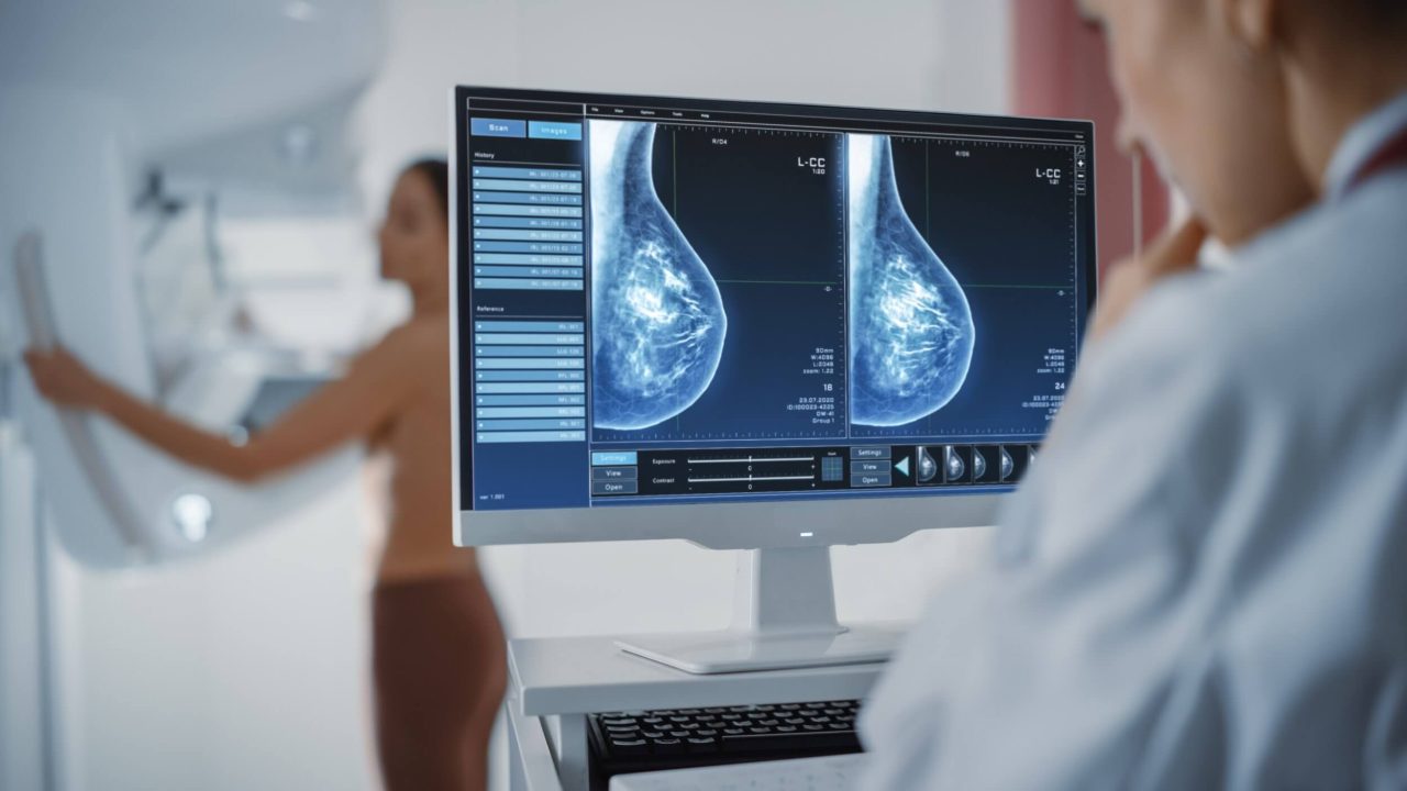
Breast mammogram. Mammograms, which is an X-ray of the breast are commonly used to screen for breast cancer. Your breast cancer specialist may recommend a diagnostic mammogram to evaluate any abnormality that is detected on a screening mammogram. Breast ultrasound. Breast ultrasound, which is more commonly used in younger women can be used to determine whether a new breast lump is a solid mass or a fluid-filled cyst. Breast magnetic resonance imaging (MRI). A breast MRI is a contrast-enhanced machine that uses strong magnets to create cross-sectional images of the breast. Confirmation of breast cancer Removing a sample of breast cells for testing (biopsy). A biopsy, where a sample of breast cells is removed for testing, is the definitive way to make a diagnosis of breast cancer. During a biopsy[2], your breast cancer specialist will use a specialised needle device to extract a core tissue from the suspicious area. Biopsy samples will be sent to a laboratory for analysis to determine whether the cells are cancerous. The pathologist will also analyse the biopsy sample to determine the type of cells involved in the breast cancer, the aggressiveness of the cancer, and whether the cancer cells have hormone receptors or other receptors that may influence your treatment options. Risk factors for Breast Cancer Leading a sedentary lifestyle or being overweight after menopause can increase your risk of getting breast cancer. Some hormone replacement therapy[3] especially those that include both estrogen and progesterone taken during menopause may also increase the risk for breast cancer if taken for more than five years. Drinking excessive alcohol and having your pregnancy after age 30 may also increase a woman’s risk of breast cancer3. The risk for breast cancer also increases with age. Most breast cancers are diagnosed after age 40[4]. A woman’s risk for breast cancer is also higher if a first-degree relative or multiple family members on the parents’ side[5] have had breast or ovarian cancer. How to prevent breast cancer Apart from regular breast screening, research shows that lifestyle changes can decrease the risk of breast cancer, even among women who are at high risk. Here are some lifestyle strategies that you can use to reduce your risk[6]: Cut down on your consumption of alcohol as this can reduce your risk of developing breast cancer. The general recommendation is to limit yourself to no more than one drink a day. Maintain a healthy body weight. Cut down on your daily calorie intake and try to incorporate some daily physical activity such as taking a walk, alighting one stop before your destination, doing housework or climbing the stairs. Breast-feeding may also play a role in breast cancer prevention. The protective effect is enhanced if you breast feed for a longer period. Limit postmenopausal hormone therapy.  Talk to your doctor about the risks and benefits of hormone therapy as you might be able to manage your symptoms with nonhormonal therapies and medications. Eating a healthy diet might decrease your risk of some types of cancer including breast cancer. Try to consume more plant-based foods, such as fruits and vegetables, whole grains, legumes, and nuts. Cut down on red meat and choose healthy fats, such as olive oil and eat more oily fish such as tuna, salmon and mackerel. References: [1]Singapore Cancer Society, Breast Cancer [2]American Cancer Society, Breast Cancer Early Detection and Diagnosis [3]Centre for Disease Control & Prevention, What Are the Risk Factors for Breast Cancer? [4]Singapore Cancer Registry Annual Report 2020 [5]Centre for Disease Control & Prevention, What Can I Do to Reduce My Risk of Breast Cancer? [6]Mayo Clinic, Breast cancer prevention: How to reduce your risk
Read more

What Does My Abnormal Mammogram Result Mean?

Most women would find getting a mammogram uneventful with an estimated 10% of women needing further follow-up tests. Every eight out of 10 women reached out for follow-up tests found no presence of breast cancer. 1 In majority of the cases, the follow-up tests find normal healthy breast tissue or benign non-cancerous breast conditions. According to Breast Screen Singapore (BSS), for every 1,000 Singaporean women who are screened for breast cancer, about 100 women are reached out for further assessment. Amongst this group of women, about 30 would be required to do a breast biopsy and about five to eight women would find that they have breast cancer. Thus, if you receive an abnormal mammogram result, it is important for you to see a doctor who can recommend the next course of action required.  The doctor may order additional imaging scans to better identify the abnormal findings. These scans can include one or more of the following: Diagnostic Mammogram This second mammogram will be more detailed and may take longer than the initial mammogram. If any of the X-ray images of your breast from the first mammogram are not clear enough, the technician will take more X- rays of specific areas of your breasts, especially the areas that are of particular concern. X-rays of specific breast areas will be taken with higher magnification or cone compression views for more detailed images to determine the presence of any breast cancer. A 3D mammogram may be done coupled with an ultrasound. Learn more about the benefits of a 3D mammogram. Breast Ultrasound The ultrasound of the breast works by picking up on the echoes produced by the high-frequency sound waves to generate an image of the tissues and the internal structures of the breasts on the computer. A breast ultrasound is a useful test for women with denser breasts. During the breast ultrasound, the technician will apply a gel on your breasts and then place a small device called a transducer on it to create images of your breast tissues. This is a painless test and it does not expose you to radiation. Magnetic Resonance Imaging (MRI) A breast MRI takes detailed images of the tissues and the internal structures of the breast. It is recommended for women who have a high risk of breast cancer, for instance, with a family history of breast cancer or presence of a hereditary breast cancer genome. After evaluating the results of these tests, your doctor will be able to advise you. Here are the three possible scenarios: 1.    The abnormal finding was nothing to worry about. Your doctor may recommend you to return to your regular screening routine. 2.    There is nothing to worry about. However, your doctor may advise you to repeat in four to six months’ time for a closer follow-up to monitor any potential changes in the breast tissues over that period of time. 3.    Breast cancer is not ruled out. The breast specialist may recommend a breast biopsy. Breast Biopsy A breast biopsy is a procedure where a small sample of tissue from your breast is removed and examined for breast cancer cells. This can be done via fine-needle aspiration biopsy, core needle biopsy, stereotactic biopsy or surgical biopsy. After the procedure, the sample tissue will be sent to the pathologist for examination. If the result returns negative or benign, no breast cancer is detected. Your breast specialist will advise you on when to have your next mammogram and whether you need any follow-up assessments. If the result returns positive and breast cancer is detected, your breast specialist will discuss treatment options with you. Received an abnormal mammogram result and not sure what is the right action to take next? Our breast specialists at Solis Breast Care and Surgery Centre will guide you every step of the way. Book an appointment with us now here. Solis Breast Care and Surgery Centre is a one-stop assessment centre that focuses on breast care and health. Our services include screening, diagnosis, procedures, surgery and post-care. In collaboration with Luma Women’s Imaging Centre, we offer bespoke breast care and women’s imaging. Solis Breast Care and Surgery Centre partners Luma Women’s Imaging Centre and offers 3D Mammograms as part of its breast screening services. Article contributed and reviewed by Solis Breast Care and Surgery Centre and Luma Women’s Imaging Centre.
Read more

Hold On – Do I Really Need A Biopsy?

Mammograms are used to detect signs of breast cancer, which is the top cancer affecting women in Singapore. Here, most mammograms are conducted in 2D, where two X-ray pictures are taken of each breast from two different angles. Sometimes, these images show breast abnormalities, but they may not be clear enough to distinguish between breast cancer and benign tumours. In cases like these, to confirm or dismiss the suspicion of breast cancer, doctors may recommend additional scans or conduct a biopsy, which is a procedure to extract a sample of breast tissue for testing. There are several types of biopsy procedures which the breast surgeon would recommend based on a few considerations such as the size and location of the suspicious area; the number of suspicious areas, and how suspicious it looks on the scan image. A core needle biopsy uses a larger gauge needle to collect samples about the size of a rice grain, while a vacuum assisted biopsy may be used to remove the abnormal area for assessment.  Sometimes, a lymph node biopsy may be required if irregularities are found under the arms. For bigger breast masses, an open surgical biopsy may be required. Most breast biopsies turn out to be benign, not cancerous. Biopsy may not be required A new technology called 3D mammography helps doctors capture multiple x-rays from different angles which are reconstructed by computer into digital images to create a 3D reconstruction of the breast. This provides clearer views of abnormalities within dense breast tissues. Greater clarity from a 3D mammogram reduces the number of false positives thereby decreasing the number of unnecessary biopsies. Be in control of your health It is important to see a doctor for further assessment if you received an abnormal mammogram report. It may not necessarily mean it is cancer. Gain clarity from a doctor on the next steps required to protect yourself against breast cancer. Solis Breast Care & Surgery Centre works with Luma Women’s Imaging to provide holistic and integrated breast care services. To increase the accuracy of the breast scan, ultrasound scans as well as a new diagnostic tool called MastoCheck can be used to help identify early-stage breast cancer. With MastoCheck, a small amount of blood is extracted to test for protein biomarkers that typically signal the presence of the disease. Solis Breast Care and Surgery Centre partners Luma Women’s Imaging Centre and offers 3D Mammograms as part of its breast screening services. Article contributed and reviewed by Solis Breast Care and Surgery Centre and Luma Women’s Imaging Centre.
Read more